Radiology Report
James Thornton
Age: 54
MRN: MRN-2026-004
MRI
Aug 27, 2026
⚠️
Demo purposes only. This AI analysis is generated by a general-purpose model and has not been trained on clinical radiology data. Results should not be used for medical decision-making.
Scan Image
Impression
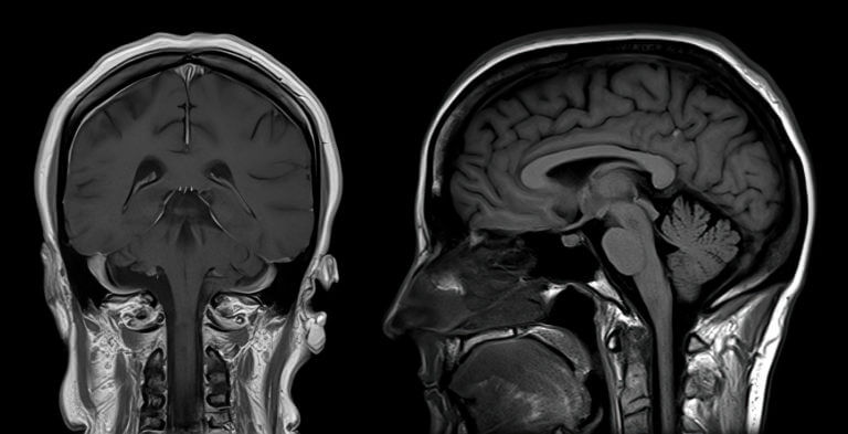
Normal brain MRI with no evidence of acute intracranial abnormality, mass effect, or hemorrhage. Age-appropriate brain parenchymal volume with preserved grey-white matter differentiation.
Findings
Brain Parenchyma — Normal grey and white matter signal intensity without focal lesions, edema, or abnormal enhancement.
Ventricular System — Lateral, third, and fourth ventricles are normal in size and configuration without hydrocephalus.
Corpus Callosum — Intact corpus callosum with normal morphology and signal characteristics on sagittal imaging.
Cerebellum and Brainstem — Normal cerebellar hemispheres, vermis, pons, and medulla without mass or signal abnormality.
Cerebral Sulci — Mild age-appropriate prominence of cerebral sulci and cortical atrophy for a 54-year-old patient.
Midline Structures — No midline shift or mass effect; pituitary gland and sella turcica appear unremarkable.
Skull and Soft Tissues — Calvarium is intact with normal bone marrow signal; visualized paranasal sinuses and mastoid air cells are clear.
Recommendation
No acute findings; routine follow-up as clinically indicated.
Model Confidence
92%
This analysis is AI-generated and intended to assist, not replace a qualified radiologist's review. Not for clinical decision-making without physician oversight.