Radiology Report
John Doe
Age: 62
MRN: MRN-2026-007
CT Scan
May 19, 2026
⚠️
Demo purposes only. This AI analysis is generated by a general-purpose model and has not been trained on clinical radiology data. Results should not be used for medical decision-making.
Scan Image
Impression
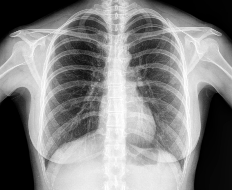
Chest radiograph demonstrates a moderate left-sided pleural effusion with adjacent atelectasis and mild cardiomegaly. No acute osseous abnormality identified.
Findings
Left pleural effusion — Moderate volume pleural effusion present in the left hemithorax with blunting of the costophrenic angle and meniscus sign.
Left basilar atelectasis — Adjacent compressive atelectasis or consolidation at the left lung base underlying the effusion.
Cardiomegaly — Cardiac silhouette is mildly enlarged with cardiothoracic ratio approaching 0.55.
Right lung — Right lung fields are clear with normal bronchovascular markings and no focal consolidation or mass.
Mediastinum — Mediastinal contours are within normal limits without widening or lymphadenopathy.
Osseous structures — Visualized ribs, clavicles, and shoulder joints demonstrate normal alignment without acute fracture or destructive lesion.
Recommendation
Recommend clinical correlation with symptoms, consideration of thoracentesis for diagnostic and therapeutic purposes, and follow-up imaging to assess interval change.
Model Confidence
88%
This analysis is AI-generated and intended to assist, not replace a qualified radiologist's review. Not for clinical decision-making without physician oversight.