Radiology Report
Linda Castellano
Age: 71
MRN: MRN-2026-005
Demo purposes only. This AI analysis is generated by a general-purpose model and has not been trained on clinical radiology data. Results should not be used for medical decision-making.
Scan Image
Impression
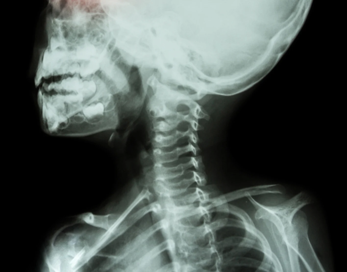
Lateral cervical spine radiograph demonstrates advanced degenerative changes throughout the cervical spine with significant anterior osteophyte formation and mild anterolisthesis at C3-C4. Underlying osteopenia is also noted.
Findings
Recommendation
Clinical correlation recommended; consider MRI if neurological symptoms are present to evaluate for spinal canal stenosis or nerve root compression given advanced degenerative changes.
Model Confidence
This analysis is AI-generated and intended to assist, not replace a qualified radiologist's review. Not for clinical decision-making without physician oversight.