Radiology Report
David Okafor
Age: 67
MRN: MRN-2026-002
Demo purposes only. This AI analysis is generated by a general-purpose model and has not been trained on clinical radiology data. Results should not be used for medical decision-making.
Scan Image
Impression
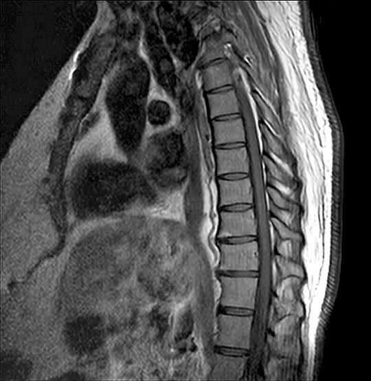
Sagittal T2-weighted MRI of the thoracolumbar spine demonstrates severe multilevel degenerative disc disease with associated anterior wedge compression deformities of multiple mid-thoracic vertebral bodies, likely representing osteoporotic compression fractures. There is marked loss of disc height and signal intensity throughout the thoracolumbar segments.
Findings
Recommendation
Correlate with clinical history of trauma, bone density studies (DEXA scan), and serum calcium/vitamin D levels. Consider short-term immobilization, pain management, and osteoporosis treatment if indicated. Neurosurgical or orthopedic spine consultation recommended for assessment of fracture stability.
Model Confidence
This analysis is AI-generated and intended to assist, not replace a qualified radiologist's review. Not for clinical decision-making without physician oversight.