Radiology Report
Sarah Mitchell
Age: 42
MRN: MRN-2026-001
Demo purposes only. This AI analysis is generated by a general-purpose model and has not been trained on clinical radiology data. Results should not be used for medical decision-making.
Scan Image
Impression
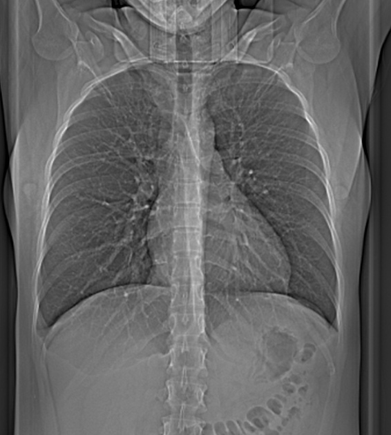
Chest radiograph demonstrates bilateral interstitial opacities with a reticular pattern throughout both lung fields, concerning for interstitial lung disease. The lungs are well expanded with no focal consolidation, pleural effusion, or pneumothorax identified.
Findings
Recommendation
High-resolution CT chest is recommended for further characterization of interstitial lung disease and to assess for fibrosis, pulmonary function tests advised, and consider rheumatologic or pulmonology consultation.
Model Confidence
This analysis is AI-generated and intended to assist, not replace a qualified radiologist's review. Not for clinical decision-making without physician oversight.