Radiology Report
Sarah Mitchell
Age: 42
MRN: MRN-2026-001
Demo purposes only. This AI analysis is generated by a general-purpose model and has not been trained on clinical radiology data. Results should not be used for medical decision-making.
Scan Image
Impression
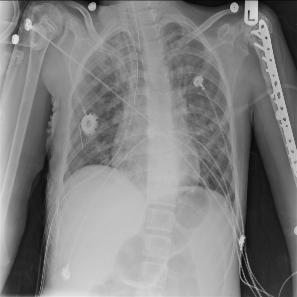
Chest radiograph demonstrates bilateral upper extremity orthopedic hardware with notable skeletal and soft tissue abnormalities, including thoracic scoliosis and rib deformities. Multiple medical lines and monitoring devices are present.
Findings
Recommendation
Clinical correlation recommended for assessment of orthopedic hardware positioning and function; follow-up imaging if clinically indicated for line placement or pulmonary concerns.
Model Confidence
This analysis is AI-generated and intended to assist, not replace a qualified radiologist's review. Not for clinical decision-making without physician oversight.